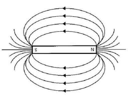
Magnetic shielding is a process that limits the coupling of a magnetic field between two locations. This can be done with a number of materials, including sheet metal, metal mesh, ionized gas, or plasma. The purpose is most often to prevent magnetic fields from interfering with electrical devices.
Magnetic Shielding For almost a century the subject of shielding extremely low-frequency (ELF) and very low-frequency (VLF) magnetic fields has been of interest 1. The interest originated from the design necessity to protect part of the circuit in radio-receiving. Magnetic shielding materials have very high permeability and 'pull' the magnetic field lines forcing them to pass through them, thus reducing the magnetic field values in the rest of the space. They are also very expensive. Materials such as copper, lead or aluminum are not suitable for shielding the magnetic fields as many people believe. Industries Served. Although our magnetic field shielding materials and products are used for low field shielding applications across a broad range of industries, Magnetic Shield Corporation provides industry specific expertise, technical engineering know-how, and design consultations to help solve increasingly high-tech advanced EMI challenges. Magnetic Shielding Magnetic lines stop here. Magnetic-field lines pass through cardboard, air, and certain other materials, depending on whether they're permeable or nonpermeable. Test different materials to see which gather magnetic lines of force and act as magnetic shields, and which allow magnetic lines of force to pass through them. Magnetic Shielding Materials are demonstrated in contact with a permanent magnet. Shielding performance is compared.Visit t.
Unlike electricity, magnetic fields cannot be blocked or insulated, which makes shielding necessary. This is explained in one of Maxwell’s Equations, del dot B = 0, which means that there are no magnetic monopoles. Therefore, magnetic field lines must terminate on the opposite pole. There is no way to block these field lines; nature will find a path to return the magnetic field lines back to an opposite pole. This means that even if a nonmagnetic object — for example, glass — is placed between the poles of a horseshoe magnet, the magnetic field will not change.
Instead of attempting to stop these magnetic field lines, magnetic shielding re-routes them around an object. This is done by surrounding the device to be shielded with a magnetic material. Magnetic permeability describes the ability of a material to be magnetized. If the material used has a greater permeability than the object inside, the magnetic field will tend to flow along this material, avoiding the objects inside. Thus, the magnetic field lines are allowed to terminate on opposite poles, but are merely redirected.
While the materials used in magnetic shielding must have a high permeability, it is important that they themselves do not develop permanent magnetization. The most effective shielding material available is mu-metal — an alloy of 77% nickel, 16% iron, 5% copper, and 2% chromium — which is then annealed in a hydrogen atmosphere to increase its permeability. As mu-metal is extremely expensive, other alloys with similar compositions are sold for shielding purposes, usually in rolls of foil.
Magnetic shielding is often employed in hospitals, where devices such as magnetic resonance imaging (MRI) equipment generate powerful magnetic flux. Shielded rooms are constructed to prevent this equipment from interfering with surrounding instruments or meters. Similar rooms are used in electron beam exposure rooms where semiconductors are made, or in research facilities using magnetic flux.
Smaller applications of magnetic shielding are common in home theater systems. Speaker magnets can distort a cathode ray tube (CRT) television picture when placed close to the set, so speakers intended for that purpose are shielded. It is also used to counter similar distortion on computer monitors.
A number of companies will custom build magnetic shields from a diagram for home or commercial applications. Shielding using superconducting magnets is being researched as a means of shielding spacecraft from cosmic radiation.
Magnetic shielding refers to the attempt to isolate or block the magnetic field of the MRI magnet. This can be done to prevent unwanted interference from the MRI magnet on nearby electronic devices. This is different from radiofrequency shielding, which is the attempt to prevent the unwanted interference of noise or radiofrequencies, often in order to avoid image distortion.


Magnetic Shielding Corp
An example of where magnetic shielding is important would be when there are nearby devices that potentially can be susceptible to magnetic interference, such as cardiac pacemakers, or other sensitive pieces of electronic or medical equipment. It also becomes relevant when low frequency external magnetic fields are being used nearby and need to be shielded from the magnetic field or fringe field from the MRI magnet. In general, the entire environment around the MR magnet needs to be protected from the fringe and magnetic fields.
A conventional marker of high magnetic field proximity is the 5 gauss line. Additional magnetic shielding may be required to protect devices that fall within the 5 G line, particularly if this extends beyond the MRI exam room. 5 G is the threshold required to avoid potential influence on pacemakers. Nearby medical imaging scanners can be influenced by magnetic field strengths as low as 1-3 G.


The methods of magnetic shielding usually involve steel or copper placed in the walls of the magnet room. These metallic steel or copper plates capture the magnetic field based on their geometric make-up and result in a cancelation or blockade of the magnetic field. The magnetic shield essentially re-directs the magnetic field so that it protects the item being shielded. One of the relevant properties of the shield is that the magnetic field is attracted to the shielding material. The amount of material in the shield is also relevant, as the more material there is, the more magnetic field it can re-direct.
Magnetic Shielding Foil
- 1. Jerrold T. Bushberg, John M. Boone. The Essential Physics of Medical Imaging. (2011) ISBN: 9780781780575
Related Radiopaedia articles
Imaging technology
- imaging physics
- x-ray
- x-ray in practice
- x-ray production
- high voltage generator
- cathode
- anode
- x-ray tubes
- filters
- grids
- x-ray film
- image intensifier
- digital radiography
- direct digital radiography
- digital image
- x-ray artifacts
- external foreign body artifact
- radiation units
- absorbed dose
- equivalent dose
- effective dose
- exposure
- legacy units
- radiation safety
- stochastic effect
- radiation-induced carcinogenesis
- stochastic effect
- radiation detectors
- dosimeters
- fluoroscopy
- computed tomography (CT)
- CT technology
- generations of CT scanners
- dual energy CT
- clinical applications of dual energy CT
- CT image reconstruction
- CT image quality
- CT dose
- CT artifacts
- patient-based artifacts
- blur
- physics-based artifacts
- hardware-based artifacts
- helical and multichannel artifacts
- MPR artifact
- helical and multichannel artifacts
- patient-based artifacts
- CT safety
- history of CT
- CT technology
- MRI
- MRI hardware
- coils
- magnets
- signal processing
- MRI pulse sequences (basics | abbreviations | parameters)
- diffusion weighted imaging (DWI)
- gradient echo sequences
- inversion recovery sequences
- perfusion-weighted imaging
- techniques
- derived values
- spin echo sequences
- MR angiography (and venography)
- non-contrast-enhanced MRA
- TRICKS
- non-contrast-enhanced MRA
- MR spectroscopy (MRS)
- 2-hydroxyglutarate peak: resonates at 2.25 ppm
- alanine peak: resonates at 1.48 ppm
- choline peak: resonates at 3.2 ppm
- citrate peak: resonates at 2.6 ppm
- creatine peak: resonates at 3.0 ppm
- functional MRI (fMRI)
- gamma-aminobutyric acid (GABA) peak: resonates at 2.2-2.4 ppm
- glutamine-glutamate peak: resonates at 2.2-2.4 ppm
- lactate peak: resonates at 1.3 ppm
- lipids peak: resonates at 1.3 ppm
- myoinositol peak: resonates at 3.5 ppm
- N-acetylaspartate (NAA) peak: resonates at 2.0 ppm
- propylene glycol peak: resonates at 1.13 ppm
- MRI artifacts
- MRI hardware and room shielding
- MRI software
- slice-overlap artifact a.k.a. cross-talk artifact
- patient and physiologic motion
- tissue heterogeneity and foreign bodies
- magnetic susceptibility artifact
- blooming artifact
- magnetic susceptibility artifact
- Fourier transform and Nyquist sampling theorem
- MRI contrast agents
- intravascular (blood pool) MRI contrast agents
- hepatobiliary MRI contrast agents
- extracellular MRI contrast agents
- contrast media safety
- MRI safety
- MRI hardware
- ultrasound
- transducers
- frame averaging (frame persistence)
- ultrasound image resolution
- imaging modes and display
- pulse-echo imaging
- Doppler imaging
- B flow
- color box
- pulse repetition frequency and scale
- color write priority
- packet size (dwell time)
- elastography
- scanning modes
- 2D ultrasound
- 4D ultrasound
- M-mode
- ultrasound artifacts
- reverberation artifact
- comet tail artifact
- hardware-related artifacts
- Doppler artifacts
- tissue vibration
- spectral broadening
- blooming
- motion (flash) artifact
- acoustic streaming
- reverberation artifact
- biological effects of ultrasound
- transducers
- nuclear medicine
- nuclear medicine physics
- nuclide
- isomer
- photopeak
- nuclide
- detectors
- scintillation detectors (gamma camera)
- emission tomography
- radiopharmaceuticals
- fundamentals of radiopharmaceuticals
- radiopharmaceutical labeling
- radiopharmaceutical production
- nuclear reactor produced radionuclides
- cyclotron produced radionuclides
- radiation detection
- dosimetry
- specific agents
- carbon-11
- chromium-51
- fluorine agents
- gallium agents
- Ga-67 citrate
- Ga-68
- iodine agents
- I-123
- I-123 iodide
- I-123 ortho-iodohippurate
- MIBG scans
- I-123 MIBG
- I-131 MIBG
- I-123
- indium agents
- In-111 Prostascint
- krypton-81m
- nitrogen-13
- oxygen-15
- phosphorus-32
- selenium-75
- technetium agents
- Tc-99m HMPAO
- Tc-99m mercaptoacetyltriglycine
- Tc-99m sulfur colloid (oral)
- xenon agents
- in vivo therapeutic agents
- nuclear medicine physics
- emerging methods in medical imaging
- radiography
- phase-contrast imaging
- CT
- deep-learning reconstruction
- ultrasound
- ultrafast Doppler imaging
- ultrasound localization microscopy
- MRI
- nuclear medicine
- total body PET system
- immuno-PET
- miscellaneous
- radiography

Magnetic Shielding Design
Promoted articles (advertising)
